
Mesonephric duct
| Mesonephric duct | |
|---|---|
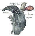
 Urogenital sinus of female human embryo of eight and a half to nine weeks old | |
| Details | |
| Carnegie stage | 11 |
| Days | 28 |
| Precursor | Intermediate mesoderm |
| Gives rise to | Vasa deferentia, seminal vesicles, epididymides, Gartner's duct |
| Identifiers | |
| Latin | ductus mesonephricus; ductus Wolffi |
| MeSH | D014928 |
| TE | duct_by_E5.6.2.0.0.0.4 E5.6.2.0.0.0.4 |
| Anatomical terminology | |
The mesonephric duct, also known as the Wolffian duct, archinephric duct, Leydig's duct or nephric duct, is a paired organ that develops in the early stages of embryonic development in humans and other mammals. It is an important structure that plays a critical role in the formation of male reproductive organs. The duct is named after Caspar Friedrich Wolff, a German physiologist and embryologist who first described it in 1759.[1]
During embryonic development, the mesonephric ducts form as a part of the urogenital system.[2]
Structure
[edit]The mesonephric duct connects the primitive kidney, the mesonephros, to the cloaca. It also serves as the primordium for male urogenital structures including the epididymides, vasa deferentia, and seminal vesicles.
Development
[edit]In both males and females, the mesonephric ducts develop into the trigone of urinary bladder, a part of the bladder wall, but the sexes differentiate in other ways during development of the urinary and reproductive organs.
Male
[edit]In a male, they develop into a system of connected organs between the efferent ducts of the testis and the prostate, namely the epididymis, the vas deferens, and the seminal vesicle. The prostate forms from the urogenital sinus and the efferent ducts form from the mesonephric tubules.
For this, it is critical that the ducts are exposed to testosterone during embryogenesis. Testosterone binds to and activates androgen receptor, affecting intracellular signals and modifying the expression of numerous genes.[3]
In the mature male, the function of this system is to store and mature sperm, and provide accessory semen fluid.The mesonephric duct (precursor of the male reproductive system) forms around the 3-4th week of pregnancy, present before the paramesonephric duct (precursor of the female reproductive system).
Female
[edit]In the female, with the absence of anti-Müllerian hormone secretion by the Sertoli cells and subsequent Müllerian apoptosis, the mesonephric ducts regress, although inclusions may persist. The vestigial epoophoron arises from these ducts. Also, lateral to the wall of the vagina, a Gartner's duct could develop as a remnant.
The mesonephric duct produces WNT9B, which is required for the elongation of the Müllerian/paramesonephric ducts (this happens before sex specification).[4]
The mesenchyme of the mesonephric duct is retained in female reproductive tissue after sexual differentiation. As the Woffian regresses, they undergo significant chromatin remodeling and move to surround the Müllerian. Cells from the Woffian mesenchyme occur in a layer around the inner epithelium, where the Müllerian mesenchyme cells also reside. The two can be distinguished by their gene expression profile: cells derived from the Woffian mesenchyme expresses AR, while those from the Müllerian mesenchyme expresses AMHR2. They appear quite evenly mixed in the oviduct, but remain clearly separated the uterus, with the Woffian mesenchyme settling in the mesometrial side.[5]
Function
[edit]Sexual differentiation
[edit]History
[edit]It is named after Caspar Friedrich Wolff who described the mesonephros and its ducts in his dissertation in 1759.[1]
Additional images
[edit]_(20754316592).jpg)
-
 Diagram of a transverse section, showing the mode of formation of the amnion in the chick
 -
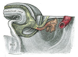
 Reconstruction of a human embryo of 17 mm
 -
 Cloaca of human embryo from twenty-five to twenty-seven days old
 -
 Tail end of human embryo thirty-two to thirty-three days old
 -
 Tail end of human embryo; from eight and a half to nine weeks old


See also
[edit]- Fetal genital development
- List of homologues of the human reproductive system
- Masculinization
- Müllerian duct
- Sexual differentiation
References
[edit]- ^ a b synd/2845 at Whonamedit?
- ^ Du, Hongling; Taylor, Hugh S. (2015). "Development of the Genital System". Principles of Developmental Genetics. pp. 487–504. doi:10.1016/b978-0-12-405945-0.00027-2. ISBN 978-0-12-405945-0.
- ^ Hannema, Sabine E.; Print, Cristin G.; Charnock-Jones, D. Stephen; Coleman, Nick; Hughes, Ieuan A. (2006). "Changes in gene expression during Wolffian duct development". Hormone Research. 65 (4): 200–209. doi:10.1159/000092408. PMID 16567946. ProQuest 1233606874.
- ^ Roly, Zahida Yesmin; Backhouse, Brendan; Cutting, Andrew; Tan, Tiong Yang; Sinclair, Andrew H.; Ayers, Katie L.; Major, Andrew T.; Smith, Craig A. (May 2018). "The cell biology and molecular genetics of Müllerian duct development". WIREs Developmental Biology. 7 (3). doi:10.1002/wdev.310.
- ^ Zhao, F; Grimm, SA; Jia, S; Yao, HH (September 2022). "Contribution of the Wolffian duct mesenchyme to the formation of the female reproductive tract". PNAS nexus. 1 (4): pgac182. doi:10.1093/pnasnexus/pgac182. PMID 36204418.
External links
[edit]- How the Body Works / Sex Development / Sexual Differentiation / Duct Differentiation - The Hospital for Sick Children (GTA - Toronto, Ontario, Canada)
| Overview | |
|---|---|
| Genetic basis |
|
| See also | |
| Authority control databases: National |
|---|